
Engineering PhD
After working as an engineer for eight years and managing to make the transition from medical device design to aerospace, I started getting the itch to apply to a graduate program. I was well positioned professionally, but I’d often wondered if I could do science and engineering in the rigorous academic setting of a Ph.D. program, and felt a need to prove to myself that I could do it.
Having seen first-hand the maturity of vehicle design at SpaceX, I chose to pursue a graduate degree focused on what I saw then as the next step—the human element of space exploration. I applied to several schools with programs in bioastronautics, visiting each campus with my husband. Depsite the distance from home and the challenges of a cross-country move, he and I both agreed that Dartmouth was clearly the best program for my goals, and in the Fall of 2019, just before the start of the pandemic, we packed up our two cats and drove across the country to New England. Thankfully I had a few months to get to know people and get a foothold before the miasma of disease, lockdowns, and remote learning descended.
During my time at Dartmouth, I’ve had the unique and valuable experience of working under two fantastic advisors in two different laboratories: Ryan Halter’s Bioengineering/Bioimpedance Lab and Jay Buckey’s Space Medicine Innovations Lab.


Dragon capsule delivering humans to the International Space Station (ISS). One of my projects is set to fly on Dragon in 2024!

Cancer cells, similar to the U-251 glioblastoma cells I used for much of my early prototyping in Ryan Halter’s lab.
Space Physiology Research
Urinary Calcium
On the space side, I have served as Principal Investigator on two NASA grants. In the first grant we developed a urinary calcium measurement device for use in spaceflight. We used a compact fluorimetric reader to detect the intensity of green light emitted by calcein-calcium complexes in sampled urine. In initial laboratory studies we refined the reagent recipe and prototyped a functional optrode. We then tested device accuracy on 100 human urine samples, enrolling participants and collecting urine per an IRB approved protocol. Each urine sample was tested by both the hospital clinical laboratory and our prototype device and agreement between the two methods was assessed with a Bland-Altman plot.

Figure 1: A) An ultra-compact device for measuring urinary calcium developed under a previous NASA HERO project. B) The generation 1 optrodes developed in the previous project. This optrode featured a solid calcein and buffer coating stabilized in an ethyl-cellulose film on the internal wall of a capillary tube. Urine was drawn into the tube inner chamber by capillary action.
Scopolamine Nebulizer
In the second grant, we are investigating the pharmacokinetics of scopolamine administered with an intranasal nebulizer. After administering the drug we perform serial blood draws in eligible participants to study the concentration of scopolamine in the blood over time, using a clinical mass spectrometer. This approach may provide astronauts relief from motion sickness at speeds comparable with IV or IM injection but without the use of needles.

Creare IN Nebulizer featuring disposable drug cartridges that could be used to administer other drugs
Numerical Modeling of the Human Cardiovascular System in Weightlessness
In addition to this NASA grant funded work, I’ve utilized a novel cranio-vascular numerical model built in MATLAB with features specific for microgravity research to study how weightlessness effects the human cardiovascular system. This modeling work has produced novel hypotheses on how the newly discovered risk of jugular venous flow thrombosis may occur in spaceflight. The model has suggested that intracranial pressure (ICP) in spaceflight is reduced rather than elevated relative to the supine position on Earth, leading our group to a novel theory of how spaceflight associated neuro-ocular syndrome (SANS) develops based solely on hydrostatic forces rather than predicated on pathologically elevated ICP. Additionally, decreased arterial pressure simulated by the model, combined with analysis of past spaceflight experiments suggests a second novel theory that could explain the paradoxical behavior of the arterial baroreflex system in microgravity, where sympathetic nerve activity is elevated while total peripheral resistance is reduced.
VARD
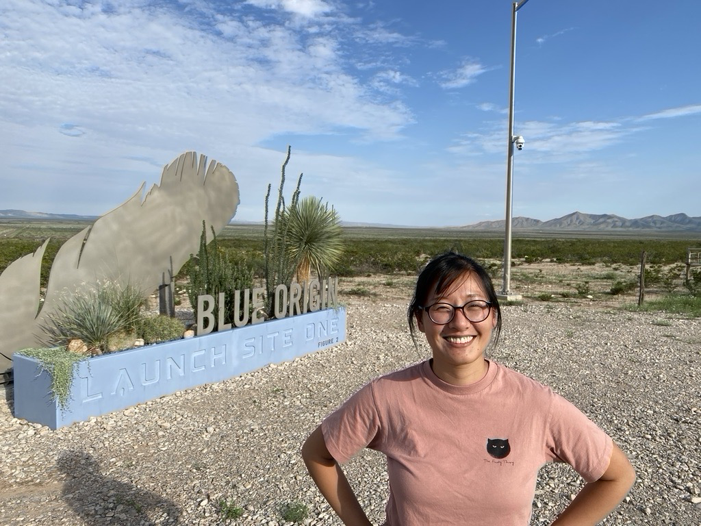
In addition to the above, my research at Dartmouth has included: assembling, programming, and managing the launch of a scientific payload that validated a microgravity compatible fluid volume sensor on a Blue Origin New Shepard flight. The sensor used the decay rate of an injected acoustic signal to determine the fluid volume in a flexible water bladder. This is technology that may be used in future EVA (extravehicular activity) suit designs in the cooling garment layer. This layer aids the temperature regulation of an astronaut’s body in the absence of convection cooling from the ambient air which is how sweat can help us thermo regulate on Earth.
Our first launch was scheduled for 2020, and after many cancellations, unsuccessful trips to Blue Origin’s launch facility in West Texas, and one gigantic explosion, it finally successfully launched in December of 2023! 🚀
One of the most valuable things I learned from this experiment was how unpredictable and logistically onerous it can be to actually fly a space experiment. Below is our timeline, complete with delays:
P10 - Scheduled for Aug 2020 launch but COVID happened.
P11 - Flight in Aug 2021. Team flew out to Texas and due to BO issues with the capsule half of the scientific payloads were demanifested (including ours)
P12 - Flight planned for Aug 2022. During launch window, launch was repeatedly scrubbed due to bad weather.
P12, second attempt - Flight was rescheduled for early Sept 2022. Rocket booster blew up and payload did not reach target altitude. Payload was recovered undamaged.
P13 - Current attempt. Was last scheduled for 10/17 but new launch date is pending.


Human Spaceflight Collaborations
I also designed a urine collection system for use on SpaceX’s upcoming Polaris Dawn Mission to study bone loss, kidney stone risk, and muscle wasting in the weightless environment, and in a collaboration with Dartmouth’s Biomedical Data Science group applied machine learning techniques to the NASA Lifetime Surveillance of Astronaut Health dataset to examine anthropometric predictors for the development of SANS.

EIT & Cell Biology
With Ryan Halter, my work has centered around a project beginning in 2019, to build a non-invasive, real-time system to track the progression of 3D tissue cultures using electrical impedance. This was funded by The Advanced Regenerative Manufacturing Institute (ARMI) in collaboration with EpiBone, a company developing technology to create bone tissue from a patient's mesenchymal stem cells in vitro, for use in autologous bone grafts. I came into this project at the start of the pandemic with almost no cell-biology or cell-culture experience and without any cell-bio expert in our lab to learn under. In the past, when finding myself in deep and unfamiliar water, I’ve found it useful to start from first principles. I took that approach here, digging through all the manuals I could find in the lab, slowly, methodically working my way through each step, designing for myself the necessary hardware (custom circuit boards, 3-D printed six well plate apparatus, bioreactors, etc.) and adapting software (algorithmic cell counters, MATLAB image reconstruction scripts, etc.).
A word here on scope: EIT is a fairly complex, technical imaging modality. Instead of getting into those details here, I’ve tried to provide a broad overview of how things evolved with this project during the last four years, focusing more on experimental design and problem solving. If you happen to be a researcher reading this and would like more detail on the specifics of my protocol or the like, please contact me—I’m more than happy to share what I have learned.
Initial Design
My first design was a simple six-well plate with electrode arrays affixed directly to the bottom surface of each well. This seemed like the most straightforward approach and this arrangement allowed me to culture a cellular monolayer directly on top of the electrode array, keeping them in close proximity to the current-driving and voltage-sensing electrodes. I began by testing which AC current frequencies and amplitudes were most acceptable to cell line I was using.

Workflow: Glue electrodes into base of well, sterilize well-electrode construct, seed cells into each well of the 6 well plate, inject current (0 - 8 mA) and wait for 48 hours, stain the cells with fluorescent dyes and incubate 30 minutes, using the microscope take images at locations of interest, count the cells in each location, determine if cell count is correlated with electric field strength.
As I carried out this initial set of experiments, a few significant problems became apparent:
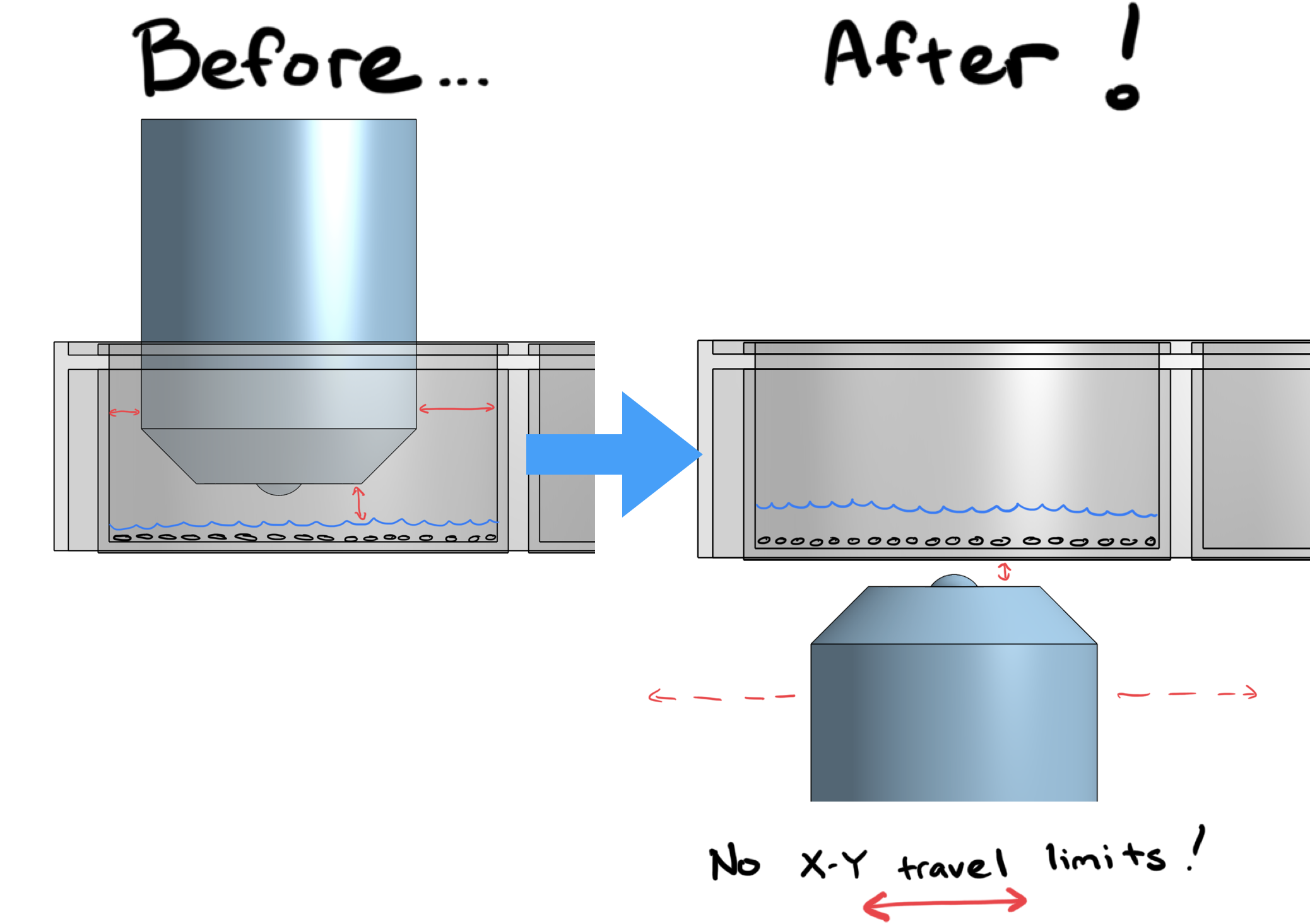
Fixing the electrodes to the base of the well obscured the view from below, which meant using an upright microscope to visualize and photograph activity in the well was my only option. However, from above, the objective lens would run into the edges of the six-well plate limiting me to a view of only a small circular area in the center of the well.
A variety of factors combined to make algorithmic cell counting difficult (electrodes in the background, cell dye/stain choice, and limitations resulting from the aforementioned upright microscopy).
Cells seemed to have trouble adhering to the flexible PCB material and the gold plated surface, making it difficult to judge if the cells were unhappy with the injected current or were absent from a region because of poor initial attachment.
Second Iteration
To address these problems, I designed and 3D printed an apparatus to suspend the electrodes above the cell monolayer in each well. Because these could be easily removed from the well at the end of an experiment, I could now use the inverted microscope to image from below, allowing the objective lens to move freely from edge to edge underneath the six well plate, with neither obstruction nor background artifact.
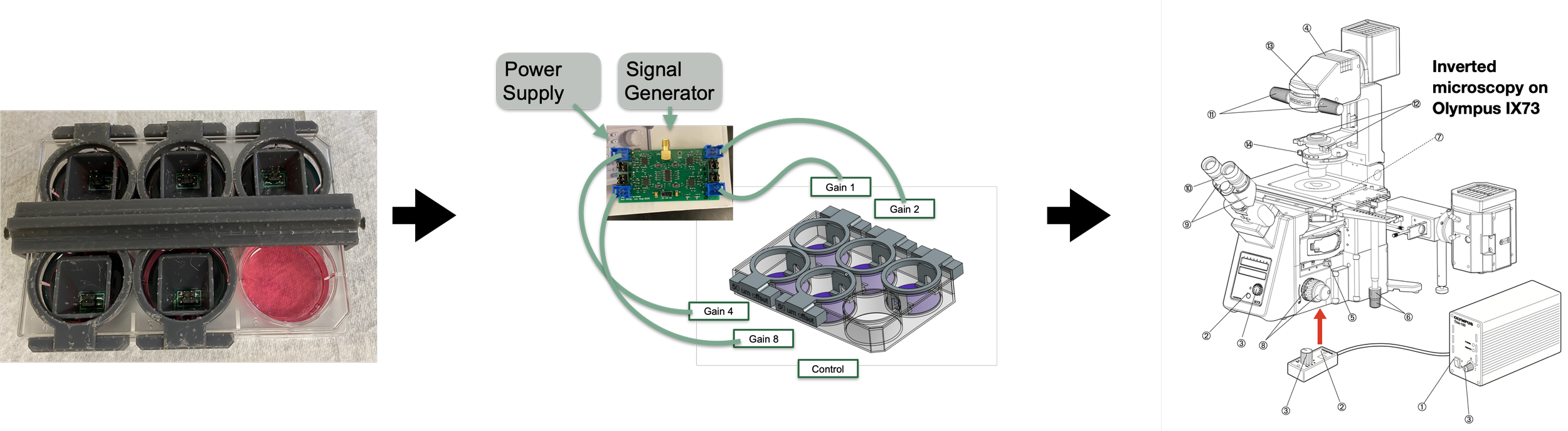
Workflow: Sterilize electrodes, seed cells into each well of the 6 well plate, after 2hr settling period, insert electrodes from above, inject current (0 - 8 mA) and wait for 48 hours, stain the cells with fluorescent dyes and incubate 30 minutes, using the microscope take images at locations of interest, count the cells in each location, see if cell count is correlated with electric field strength.
Problems Solved
🥳
Problems Solved 🥳
Objective Lens Collisions
With the upright microscope, the objective lens would run into the edges of the six-well plate, producing images with a limited view of the small circular area in the center of the well. My removable, suspended electrode-array made the cells visible through the transparent bottom of the six-well plate, allowing me to image with the inverted microscope, free of obstruction and artifact.

Cell Adherence
In my initial design, it often seemed like the cells had trouble adhering to the flexible PCB material and the gold plated surface, making it difficult to judge if the cells were unhappy with the injected current or were absent from a region because of poor initial attachment. By suspending the electrode array above the cellular monolayer, the cells could grow happily on the bottom of well, allowing me to isolate the effects of various AC current and frequency combinations on cell growth.

Automated Cell Counting
Initially I had used a Live/Dead cell stain (Calcein/EthD-1), before switching to Calcein only which yielded marginally better (but still tough to automate) results. To make it easier for the algorithm to pick out cells accurately, I decided to stain the cell bodies and nuclei separately. This also allowed me to tune the two images with software before combining them into a composite image, which made counting the cells manually much more reliably accurate, if still time consuming and tedious. However, by comparing a number of manual counts to algorithmic counting of the Hoescht Channel images, I was able to validate the accuracy of the cell-counting algorithm.

“We have never arrived. We are in a constant state of becoming.”

Moving to 3D Tissue Cultures
This design was a significant improvement, but as my experimental needs grew more complex and I began experimenting on 3D tissue cultures, I needed to access the well to change media twice a week. Unfortunately, removing and replacing the electrode arrays to replace the media created small positional differences that interfered with the generation of high quality time difference impedance data, and this issue proved difficult to mitigate. Additionally, this process would inadvertently create air bubbles that collected on the electrode surface, creating high-impedance artifacts in the data. I needed a repeatable, reliable, but still not-too-tedious way to lock everything in at the start of an experiment and perform the media change without disturbing the electrodes and cell culture scaffold. After some literature review and careful planning, I set my sights on creating a bioreactor with good visual and physical access.

Third Iteration: Bioreactor
My bioreactor design centered around a 3D printed cell-culture chamber where my rigid electrode array PCB could form one water tight wall of the chamber and a clear cover plate formed the other water tight wall. The 3D printed chamber contained ports to allow media to flow through, and male luer threads on either side to easily connect to 1/4 inch silicone tubing. The clear cover plates were originally laser cut from acrylic sheets for convenience, but I soon switched to polycarbonate once I discovered (on a critical experiment day) that acrylic had a tendency to crack when it came in contact with alcohol.


ARMI Live Technology Demo
For this demo, I injected conductive solution bolus (blue) and insulating solution bolus (red) into the chamber and could visualize the conductivity change in realtime in Matlab. I used OBS Studio for demo, which made it look extra polished.
Once I had the bioreactor chamber designed, I had initially planned to slowly push cell media through with a syringe pump, but switched to a peristaltic pump with an air permeable media bag, after learning the importance of gas exchange in closed loop culture systems.


After many iterations I now have a fully-functioning custom designed and fabricated bio-reactor system, which integrates our lab’s specialized electrical impedance tomography hardware. I am continuing to iterate on this design, working toward a system that can non-destructively characterize human cell-growth and differentiation in real time.

-
Buckey, J. C., Thamer, S., & Lan, M. (2023). Bone loss and kidney stone risk in weightlessness. Current Opinion in Nephrology and Hypertension, 32(2), 172-176.
Lan, M. et al. Proposed mechanism for reduced jugular vein flow in microgravity. Physiological Reports 9, e14782 (2021).
Buckey, J. C., Lan, M., Phillips, S. D., Archambault-Leger, V. & Fellows, A. M. A theory for why the spaceflight-associated neuro-ocular syndrome develops. J Appl Physiol 132, 1201–1203 (2022).
Buckey, J. C. & Lan, M. Why central venous pressure falls below supine levels in weightlessness. J Appl Physiol (2022) doi:10.1152/japplphysiol.00271.2022.
Lan, M., Akin, M. V., Anderson, A. & Buckey, J. C. Microgravity-induced reduced jugular vein flow is more pronounced on the non-dominant side. Acta Astronaut (2022) doi:10.1016/j.actaastro.2022.05.048. -
Aerospace Medical Association, Chicago, May 2024 – Extreme Reductions in Body Mass Index as an Analog for Weightlessness to Examine Volumetric Changes in the Brain
• Aerospace Medical Association, Chicago, May 2024 – Nebulized intranasal scopolamine may provide rapid relief for space motion sickness.
• NASA HRP IWS, February 2024 – Ultra-Compact Urinary Calcium Measurement Device: Refinement and Application, Final Data
• NASA HRP IWS, February 2024 – Nebulized intranasal scopolamine may provide rapid relief for space motion sickness.
• NASA HRP IWS, February 2024 – Eye length is a potential predictor of spaceflight- related visual changes
• NASA HRP IWS, February 2024 – A case definition is needed for the Spaceflight Associated Neuro-ocular Syndrome.
• International Astronautical Congress, Baku, October 2023 – A case definition is needed for the Spaceflight Associated Neuro-ocular Syndrome.
• International Astronautical Congress, Baku, October 2023 – Eye length is a potential predictor of spaceflight-related visual changes
• International Space Station Research and Development Conference, Seattle, August 2023 – Technology demonstration of Ultra-Compact Urinary Calcium Measurement Device
• Advanced Regenerative Manufacturing Institute, Manchester NH, June 2023 – Technology demonstration of electrical impedance tomography for monitoring cell growth on a decellularized bone scaffold.
• Aerospace Medical Association, New Orleans, LA, May 2023 – Ultra-Compact Urinary Calcium Measurement Device: Refinement and Application
• Aerospace Medical Association, New Orleans, LA, May 2023 – A re-evaluation of the acute effects of weightlessness
• BMES ABioM SIG, Hyattsville, MD, March 2023 – Current injection amplitude and frequency vs cell count for non-invasive Electrical Impedance Tomography applications
• NASA HRP IWS, February 2023 – Ultra-Compact Urinary Calcium Measurement Device: Refinement and Application
• NASA HRP IWS, February 2023 – The Acute Effects of Head Down Tilt and Weightlessness Differ Fundamentally
• NASA HRP IWS, February 2023 – Why the Spaceflight-Associated Neuro-Ocular Syndrome Develops: A Theory
• NASA HRP IWS, February 2023 – Reduced Intrathoracic Pressure Does Not Explain the Reduction of Central Venous Pressure Below Supine Levels in Space
• International Astronautical Congress, Paris, September 2022 – A theory for unexplained vasodilation with elevated noradrenaline levels in spaceflight.
• Advanced Regenerative Manufacturing Institute, Manchester NH, June 2022 – Electrical impedance tomography for monitoring cell growth on a decellularized bone scaffold.
• Aerospace Medical Association, Reno NV, May 2022 – Lower body negative pressure may not be a suitable countermeasure for spaceflight associated neuro-ocular syndrome.
• NASA HRP IWS, February 2022 – Updates to the ultra-compact device for monitoring bone loss and kidney stone risk.
• International Astronautical Congress, October 2021 – Microgravity-induced reduced jugular vein flow is more pronounced on the non-dominant side.
• International Conference on Environmental Systems, July 2021 – Simulated cardiovascular responses in microgravity and head-down tilt.
• NASA HRP IWS, February 2021 – Proposed mechanism for reduced jugular vein flow in microgravity.
• NASA HRP IWS, February 2021 – Ultra-compact device for monitoring bone loss and kidney stone risk.
-
Title: Nebulized intranasal scopolamine for rapid treatment of motion sickness
Role: Co-PI
Major Goals: The major goal of the project is to evaluate the pharmacokinetics of nebulized scopolamine administered intranasally to verify that rapid onset is achieved.
Status of Support: Active
Project Number: AWD00012329
Name of PD/PI: Lan
Source of Support: NASA Omnibus
Title: Ultra-compact urinary calcium measurement device: refinement and application
Role: PI
Major Goals: The major goal of the project is to refine the accuracy, precision, and ease of use for a technology that measures urinary calcium levels in spaceflight.
Status of Support: Active
2
Mimi Lan • Phone: (949) 798-9247 • E-mail: mimi.lan.th@dartmouth.edu
Project Number: 80NSSC22K0847 Name of PD/PI: Lan
Source of Support: NASA Omnibus
Title: Kidney stone, bone loss, and muscle atrophy risk on short duration flights
Role: Graduate student
Major Goals: The major goal of this project is to collect first morning urine void on a SpaceX civilian spaceflight and obtain measures of kidney stone risk and muscle atrophy.
Status of Support: Active
Project Number: 32575 (Rapport)
Name of PD/PI: Buckey
Source of Support: Bisu, Inc
Title: Volume Sensor for Flexible Fluid Reservoirs in Microgravity
Role: Graduate student
Major Goals: The major goal of the project is to develop a novel sensor that detects the remaining volume of coolant water stored in a flexible bladder in the advanced space suit. This technology will be validated on a Blue Origin New Shepard Payload flight.
Status of Support: Active
Project Number: 80NSSC18C0223/S627
Name of PD/PI: Phillips
Source of Support: NASA / Creare, LLC (Prime)
Title: Monitoring Tissue Growth Attributes with Bioimpedance Imaging
Role: Graduate student
Major Goals: Use minimally-invasive, low-power, small-form-factor electrical impedance technology to provide real-time data regarding the growth attributes of 3D tissue culture. Status of Support: Active
Project Number: W911NF-17-3-0003
Name of PD/PI: Halter
Source of Support: Advanced Regenerative Manufacturing Institute, Inc.
Title: Ultra-compact device for monitoring bone loss and kidney stone risk
Role: Graduate student
Major Goals: The major goal of this project is to develop and validate a novel, practical technology for measuring urinary calcium levels in flight.
Status of Support: Completed
Project Number: 80NSSC19K1632
Name of PD/PI: Buckey
Source of Support: NASA Omnibus